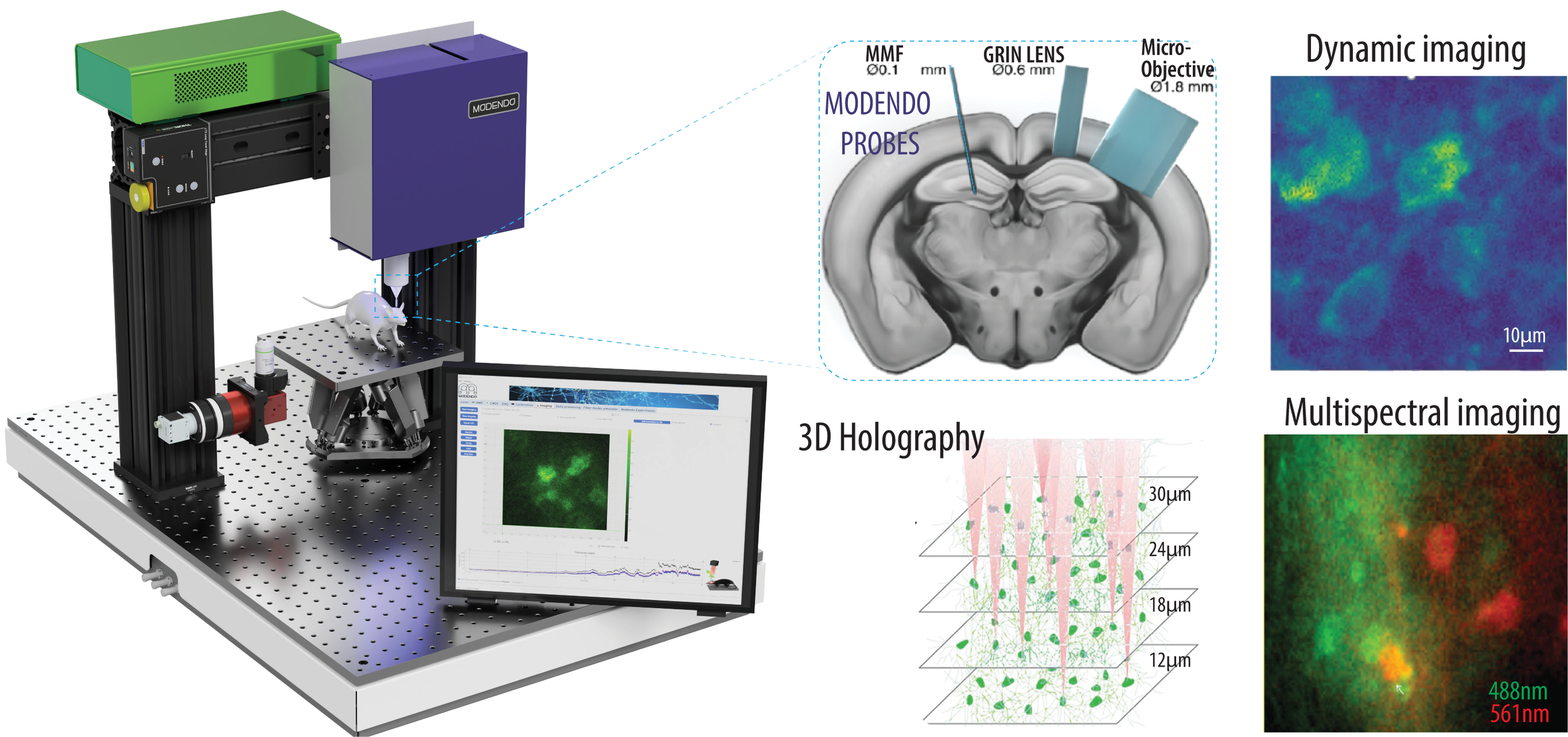
Minimally invasive imaging in the insular cortex and other subcortical areas involved in taste and reward.
Fiber optic imaging of taste-responsive neurons in the VPMpc. (A) graphic of behavioral set up and integration with Modendo’s NeuroVu Prototype. (B) Sample trace from a Soma-GCaMP7f expressing neuron during presentation of 3 tastants.
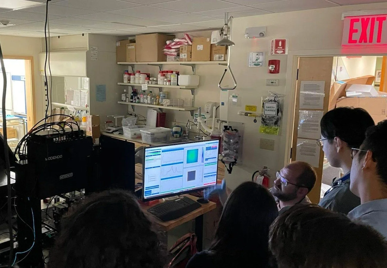
Imaging Structure and Function in the Nervous System course
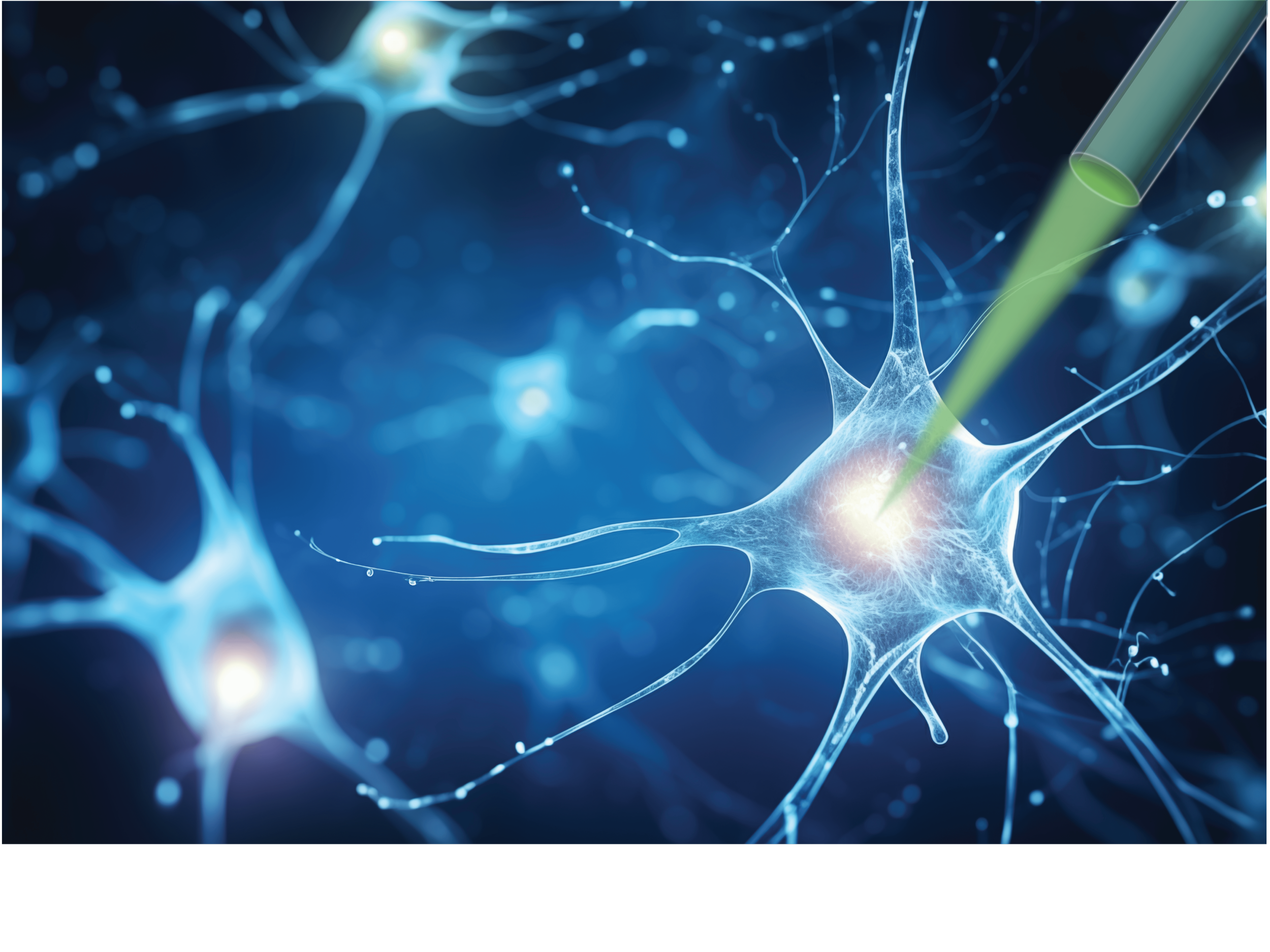
Cell and circuit-specific dynamic recordings within the rodent brain.
Live imaging of GCaMP+ neurons as the fiber optic probe is lowered into the striatum. (Left) 100 micron diamater fiber optic probe moving in z(depth) during fluorescent imaging (488nm). (Right) Experiments performed at CSH 8/25. Multiple GCaMP sensors were tested as well as voltage sensors.
Ana Clara Bobadilla Research Lab
Tracking and tuning of cells within deep drug-specific neuronal ensembles, employing multispectral imaging and complex time-locked behavioral cues.
Ultrathin endomicroscopy allows for the detection of unique neural ensembles in deep brain regions. TD-Tomato+ cells were tagged after drug exposure and co-infected with CA2+ reporters (not shown).
Dan LaBarbera Research Lab
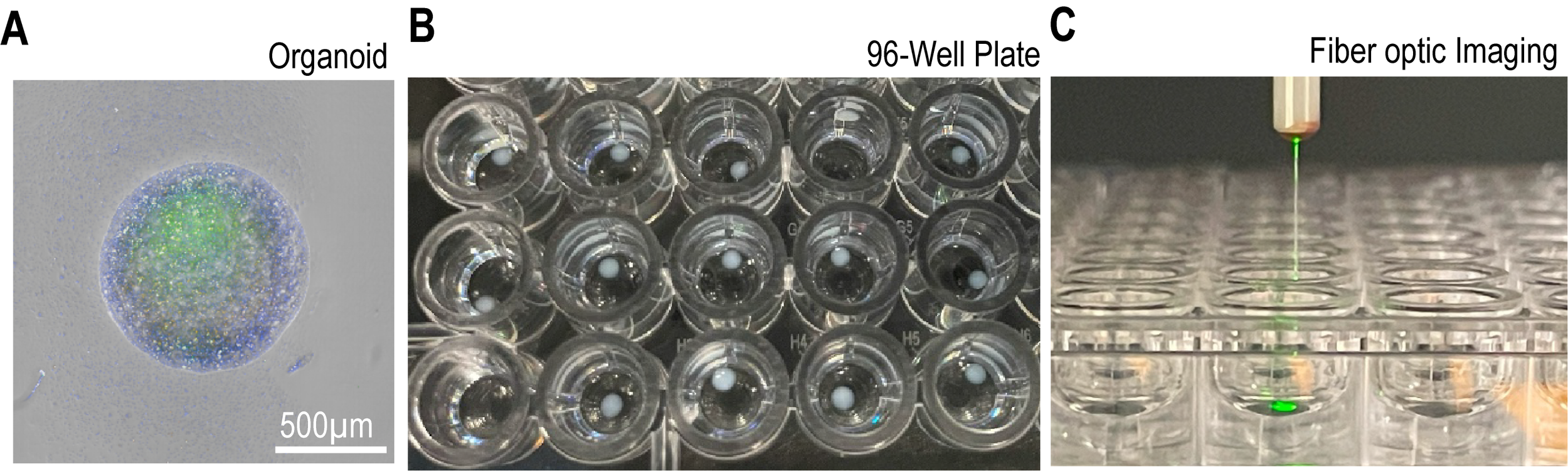
Fiber optic imaging of organoids in cell culture with a future focus on pharmaceutical testing and modeling in the context of personalized medicine.
In Vitro imaging of 3D-printed Osteosarcoma organoids. (A) Rep. image of a single organoid using a conventional confocal (E-Cadherin-488nm) (B) Organoids were 3D printed into 96-well plate, 1mm thick. (C) Rapid 3D imaging using Modendo probe.
Laboratory of Applied Photonics Devices (LAPD)
Advancements in optical imaging- FLIM- and clinical applications within cancer.
FLIM imaging in Switzerland with Modendo’s prototype. Researchers will use this tehcnology to image ex vivo tissue from tumor biopsy in head and neck cancer.
Modendo In-Vivo Research Lab
Advancements in in vivo ultrathin endomicroscopy: Multi-site imaging, optogenetics, multispectral, dynamic imaging (GCaMP) and photoablation.