
Neuroscience Applications
See deeper.
Preserve the biology.
Modendo helps researchers image and stimulate neural activity deep within the brain through an ultrathin probe designed to minimize tissue disruption.
THE CHALLENGE
The closer you get, the more your tool can change what you observe.
Deep-brain imaging often requires probes large enough to damage tissue, disrupt blood vessels, or alter the neural environment being studied.
That creates a difficult tradeoff: reach the cells that matter or preserve the biological system needed to understand them.
THE MODENDO SOLUTION
A microscope built around the biology.
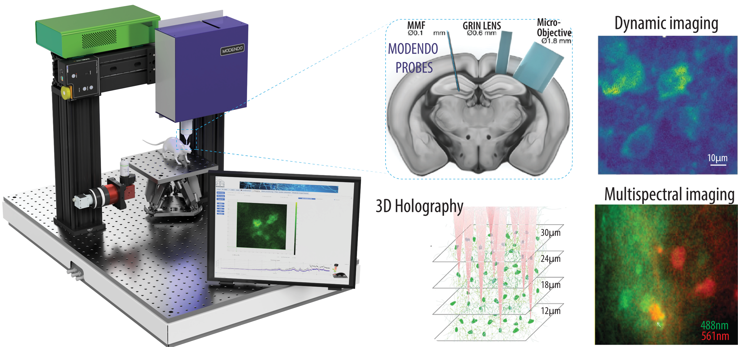
Modendo controls light through fiber probes ranging from approximately 60 to 200 µm in diameter.
The same probe can deliver illumination, collect fluorescence, and project targeted stimulation patterns—providing cellular-level access to deep brain regions with a significantly smaller physical footprint.
Accessing deeper value for your team.
For Principal Investigators
Ask new questions in deep and difficult-to-access brain regions while protecting the integrity of the experimental model.
For Researchers and System Users
Combine real-time fluorescence imaging, multispectral acquisition, volumetric scanning, and photostimulation in one adaptable system.
For Core Facilities and Research Partners
Offer a differentiated imaging capability that supports new collaborations, experimental designs, and application-specific configurations.
USE CASES
Built for the questions conventional probes cannot answer.
-
Visualize cells and neural activity in subcortical regions through an ultrathin optical path.
-
Record GCaMP and other optical indicators as proxies for neural activity in real time.
-
Track identified cell populations across behavioral events, sensory cues, or pharmacological conditions.
-
Use multiple wavelengths to distinguish signals, cell populations, or reporters within the same experiment.
-
Project controlled light patterns at the probe tip for optogenetics and other light-driven interventions.
-
Scan multiple focal planes to reconstruct high-resolution three-dimensional information.
IMAGING RESULTS
See the biology, not the disruption.
Live Imaging in the Striatum
Video of GCaMP-positive neurons as a 100 µm fiber probe moves through depth during fluorescence imaging.
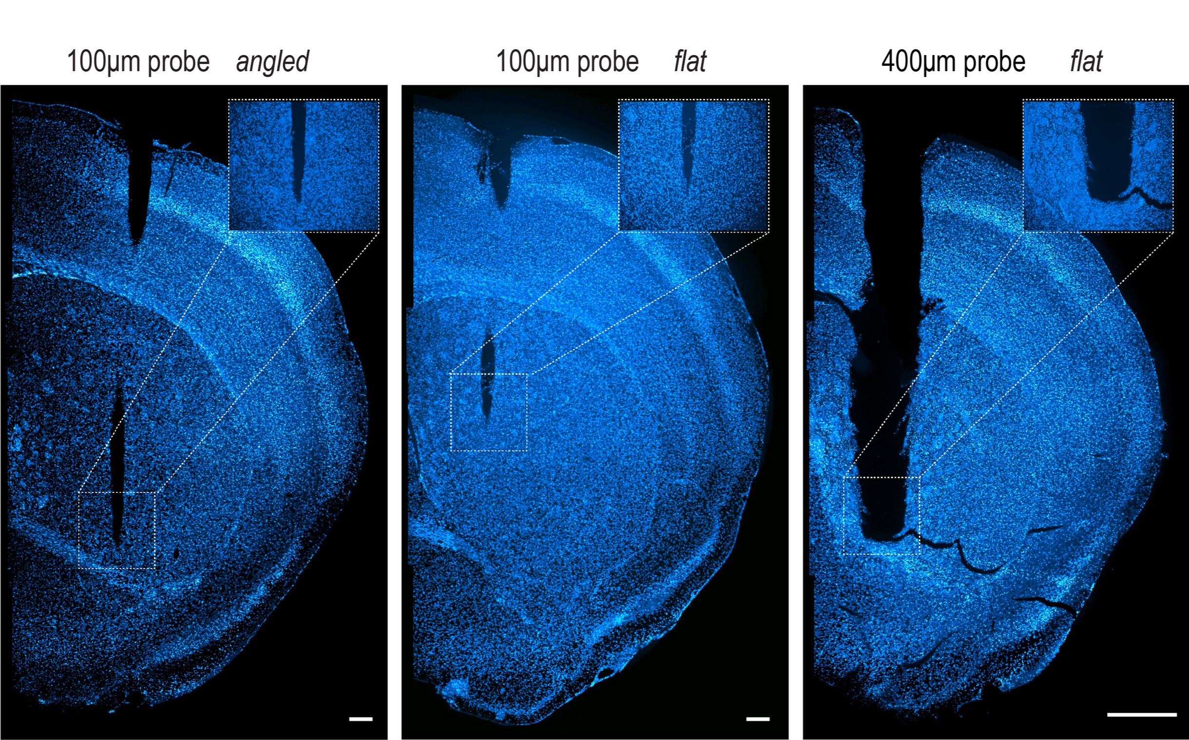
Tissue-Impact Comparison
A visual comparison between Modendo’s ultrathin probes and larger probes commonly used for photometry.
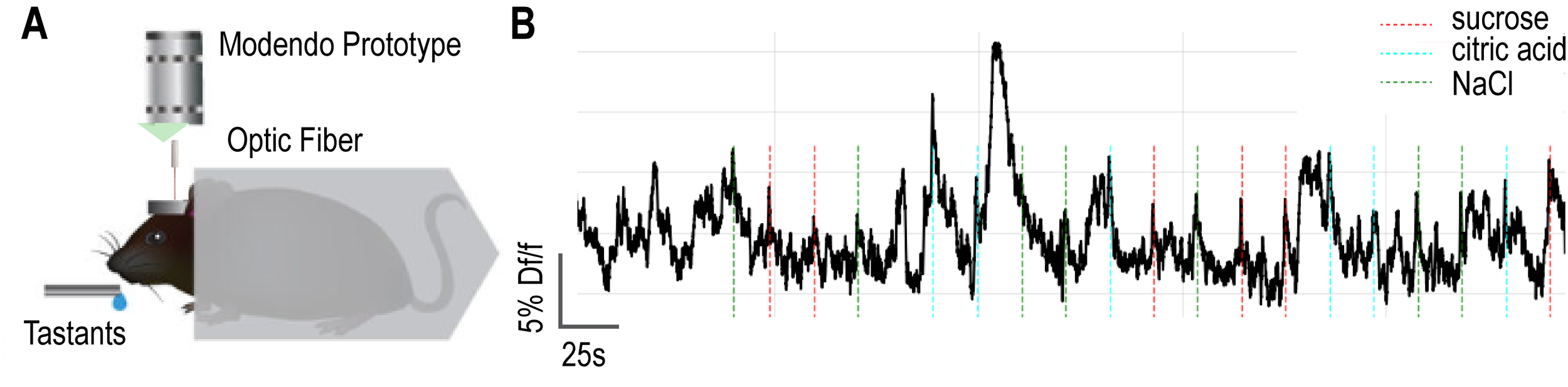
Neural Activity in Deep Regions
Representative images and activity traces from research collaborations studying taste, reward, and drug-associated neural ensembles.
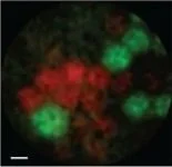
Multiplane and Multispectral Results
Imaging examples demonstrating depth scanning and simultaneous optical signals.
TECHNICAL CAPABILITIES
Designed for demanding in-vivo research.
Probe diameters from approximately 60 to 200 µm
High-resolution cellular imaging
Real-time in-vivo fluorescence imaging
Illumination and collection through the same probe
Multiple excitation wavelengths
Multispectral imaging
Three-dimensional focal-plane scanning
Computational and machine-learning reconstruction
Neural activity detection and sorting
Arbitrary-pattern projection for photostimulation
No moving scanning components at the probe tip
Application-specific probe terminations
Tell us what you’re trying to observe.
Share the brain region, optical signal, and experimental question. We’ll explore how Modendo can support the study while minimizing its impact on the biology.